100 Jahre MPI

DE
  • Welcome
  • Psychiatry and Neurobiology
  • Imaging methods
  • Anatomy
  • Neurophysiology
  • Molecules
  • The impact of genes
  • Epigenetics
  • Research methods
  • Therapies of tomorrow
  • Genetics of psychiatric disorders
  • Animal models
  • Bringing new things to light
  • Psychiatry and Neurobiology

    Imaging methods

    Anatomy

    Neurophysiology

    Molecules

    The impact of genes

    Epigenetics

    Research methods

    Therapies of tomorrow

    Genetics of psychiatric disorders

    Animal models

    Bringing new things to light

  • Psychiatry and Neurobiology
    100 years of research and therapy The 100 years of the Max Planck Institutes of Psychiatry and Neurobiology reflect the development of psychiatry in the 20th century in Germany: From the mere observation of people with behavioral problems to the molecular genetical dissection of the brain. Emil Kraepelin, founder of the predecessor institute, created a classification of psychiatric diseases, which is still valid to this day. He meticulously logged the symptoms of each patient on observation sheets. With this, a scientifically based psychiatry was born. More and more diagnoses were added over the following decades. Today, the worldwide available diagnostic catalogue DSM V lists 374 commonly known psychiatric diseases. However, the higher number of diagnoses was not matched by a higher number of therapeutical possibilities. Today, scientists follow a different approach. It is now known that diagnoses do not necessarily correspond to the affected biological mechanisms in the patient’s brain. Patients with depression, for example, can present with different pathophysiological processes. Although these patients would profit from distinct therapies, they currently all receive the same diagnosis and are therefore treated uniformly. Conversely, neuroscientists have shown that one altered mechanism in the brain can cause various symptoms, which would result in distinct diagnoses. Here, patients receive diverse therapies, even though they could profit from one and the same treatment. Crucial advances in psychiatry will therefore depend on personalized, scientifically based medicine.
  • Imaging methods
    Mapping the living brain shows the brain’s activity By applying a staining method developed by Franz Nissl, Korbinian Brodmann examined histological sections from human brain tissue to develop the so-called Brain Atlas. It describes a total of 52 areas which were postulated to perform different functions. This anatomical map is valid to this day. In 1919, a new technique based upon the application of X-rays (pneumencephalography) provided an insight into the brain of living patients. This highly invasive procedure was replaced in the 1970s by the Computer Tomography (CT), which provided for the first time virtual sections in a non-invasive manner. The clinical break-through in Neuroimaging occurred in the 1980s with the introduction of Magnetic Resonance Imaging (MRI). This technique allows for the acquisition of images from the patient’s brain in high resolution and contrast, without causing any side effects. Moreover, with the help of the MRI, it is possible to extract information with respect to the brain metabolism, microstructure and functionality. Thus, it is possible today to characterize in detail the metabolic processes, brain region connectivities and activity of the living brain. Out of this multitude of information disease specific and image based features of psychiatric disorders can be identified, helping us to optimize diagnosis and therapy.
  • Anatomy
    Details of form, structure and position become visible In the early 1900s, novel staining methods such as the Nissl and Golgi stains, enabled the identification of individual nerve cells and the investigation of their shape and position in thinly cut slice of brain. The discovery of fluorescent dyes and improvement of microscopy tech- nology made it possible to label nerve cells in living brain tissue and study their activity, in addition to their anatomy. This facilitated, for example, the better understanding of microglia cells, synaptic plasticity during learning and memory, and the motion vision of flies. Even finer details are visible using enhanced electron microscopy, which is now able to show, at high resolution, the neuronal structures and connections of the brain in three dimensions: The generation of a complete circuit diagram of the brain is finally within reach.
  • Neurophysiology
    Micro-electrodes and new methods reveal biochemical processes of nerve cells Nerve cells communicate using electrical signals generated by the selective movement of ions through the cell membrane. With the aim of understanding these processes better, the discipline of electrophysiology was born in the early 20th century. Wolfram electrodes placed within the extracellular space showed voltage changes and thus nerve cell activity. The development of sharp (glass) electrodes enabled measurements within individual cortical nerve cells. Aside from numerous insights into the functioning of nerve cells, pioneers of electrophysiology such as Otto Creutzfeld and Hans Dieter Lux were able to show that the electrical signals observed on the skull surface during an electroencephalogram (EEG) originate from synaptic activity of numerous nerve cells in the cortex. Erwin Neher and Bert Sakmann later developed in Göttingen the patch-clamp technique: A method able to measure the electrical current through a single ion channel in the membrane of living cells (for which they received the Nobel prize in 1991). Today, patch-clamp is one of the most important neurophysiological techniques, it even enables the measurement of electrical activity of individual nerve cells in the fruit fly brain. Remarkably, dyes can now be used to study the point of origin and distribution of single action potentials and other physiological processes non-invasively under the microscope.
  • Molecules
    The role of cell components In order to understand biological mechanisms underlying disease, a detailed analysis and delineation of their molecular components are mandatory. After the completion of the human genome project, the identification of genes causally linked to disease has made great progress. However, the 20,000 plus genes are only the beginning. When and where they are expressed in cells and tissues is controlled by a multitude of other molecular entities including micro RNA, transcription factors, histones and epigenetic mechanisms including covalent modifications of the DNA itself. Proteins and metabolites are the functional molecules: They determine cellular activity and are involved in a great number of processes including signal transduction, metabolism and others. In order to develop therapies and medicines, we require an understanding of the cellular and molecular processes in health and disease. Unlike in other areas of medicine, there is currently no blood test that the psychiatrist can use for diagnosis or monitoring treatment response. Dynamic molecular biosignatures that reflect disease state and drug response will eventually complement current clinical diagnosis and enable precision psychiatry.
  • The impact of genes
    Insight into physiological processes and disease mechanisms The most important area of genetic research is molecular genetics, investigating the molecular principles of inheritance. The main focus of molecular genetics lies on traits of genetic information (generally DNA), DNA replication and ensuing mutations – which can lead to new varieties and disorders, and the decoding of genetic information into RNA and proteins. Today, scientists are able to target single genes and modify or silence them, for example with the knock-in/knock-out and genome editing technologies. This makes it possible to study only a few specific nerve cells in the intact brain of laboratory animals, to manipulate these cells and study their contribution to a specific behavior. Genetic tools can identify molecules and study their role during the development of the nervous system, in developmental disorders, neurodegenerative diseases or age related processes. The aim of these studies is to better understand the basic molecular mechanisms, which could eventually lead to the development of new therapeutic approaches.
  • Epigenetics
    How the environment changes the genome Stress and trauma are risk factors for psychiatric diseases. These environmental experiences can directly affect our genome and thereby also reprogram our nerve cells. Environmental influences can thus cause long lasting changes in our ability to react to stress, which in turn increases the susceptibility for psychiatric disorders. The genome can be affected by so called epigenetic changes. In contrast to genetic alterations, epigenetic changes do not impair the sequence of single DNA components, the bases, but rather influence the efficacy with which the information coded by the DNA sequence is read. Epigenetic alterations caused by environmental experiences are DNA methylation and histone modifications (fig.). These alterations result in more or less tightly packaged DNA, which in turn impacts the accessibility of distinct proteins to certain genes. An additional mode in which the environment can exert influence on the genome is through changes in the amount of so called micro RNAs. These molecules can interfere with the protein production process. Genetic predisposition can contribute considerably to the role of epigenetic changes in many psychiatric diseases. A better understanding of the processes involved could help to reverse the effects of negative environmental experiences and thereby improve treatment of psychiatric diseases or prevent them from developing in the first place.
  • Research methods
    Where do we go from here? Genetic and biochemical processes underlying the development of neurological, neurodegenerative and psychiatric diseases require investigation at the preclinical level. Recent advances in the field of genetic engineering and the very high similarity between the human and the mouse genome have provided scientists with the ability to generate and establish mouse models (preclinical models) for a large variety of genetic-based brain disorders. New techniques and systems are being developed in order to better link the assessed parameters in the animal models with the human conditions. These techniques involve continuous measurement of both behavioral and physiological data over many days in a semi-natural environment and in a group of mice. Thus, the animal models of disease provide robust and accurate approaches to measure the cause and effect of various interventions and to test efficacy of potential therapeutic treatments. Similarly, patient-derived so called cerebral organoids or “mini-brains” hold promise for regenerative medicine, testing of drug responses and the understanding of molecular and cellular properties of the patient’s brain. Organoids are generated from generic cell samples of the donor, such as blood- or skin cells. These cells are reprogrammed to grow from a naïve state to a 3D complex in vitro structure consisting of brain-specific cell types. The structures recapitulate in a very simplified manner the development of the human brain, more specifically the patient’s brain. Due to the simple production of and the close resemblance to human organs, organoids may open new avenues for translational research and invite an almost immediate application into the clinic.
  • Therapies of tomorrow
    Personalized medicine should lead to new treatments In the last decade, research advances in imaging and molecular genetics have provided new insights into the complex developmental mechanisms underlying psychiatric disorders such as schizophrenia, depression and bipolar disorder. These advances have provided the basis for a more precise classification of disease and the definition of neurobiologically founded subgroups. As a consequence, new therapeutic approaches are arising. One example is the gene FKBP5: It is essential for a correctly functioning stress response. Some variants of the gene increase the stress response beyond their normal level and thus increase the risk of psychiatric disorders. If these gene variants are detected in patients, medication could be given to block the overactivity of this gene. By considering all characteristics of individual patients, a customized therapy can be provided, which is quicker to work and more efficient. With the help of personalized medicine, new biomarkers will be discovered, which will allow us to predict the risk of a disease occurring and its progression. Psychotherapy is a fundamental component in the treatment of all psychiatric disorders. Here as well, the molecular, biochemical and cellular mechanisms are being investigated in order to better assess the appropriate therapy for individual patients.
  • Genetics of psychiatric disorders
    Although psychiatric disorders can be more frequent in some families than others, they do not simply follow a predictable inheritance pattern The following mechanisms are known to contribute: Rare gene variants Genetic alterations can occur spontaneously in the affected individual and are not found in the parents. In most cases, these alterations represent rare genetic changes in which entire gene sections are deleted or duplicated (copy number variants). These increase the risk for e.g., schizophrenic disorders and autism. Gene variants are not deterministic Although affected patients carry risk variants, they can also be found in unaffected people. Many risk variants occur commonly in the population and are therefore present in many healthy individuals. Polygenetic heredity Psychiatric diseases in particular result from a summation of many risk variants. Only in rare cases is the trigger factor for a psychiatric disease a single gene variant. Genetics and environment Many gene variants only become risk factors in the context of specific environments, such as stress. Within a positive environment, the same gene variant can turn out to be advantageous. Psychiatric diseases arise from a complex combination of a multitude (hundreds to thousands) of gene variants and environmental influences at distinct developmental stages – from fertilization to old age.
  • Animal models

    Basic research broadens knowledge. It is only when components and functions of an organ, tissue or cell group are known and understood that approaches for an effective therapy with minimal side-effects can be developed.
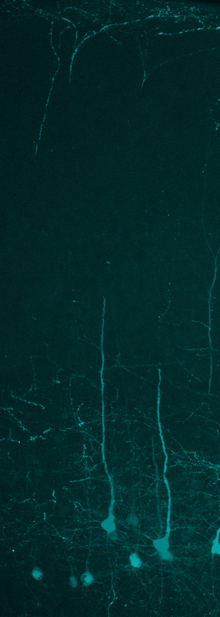
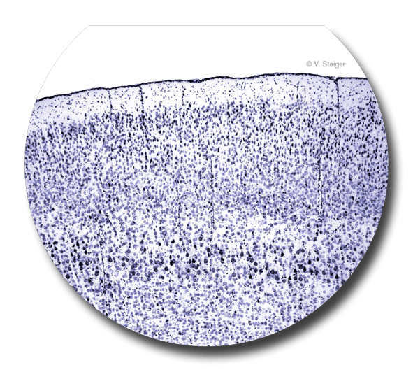
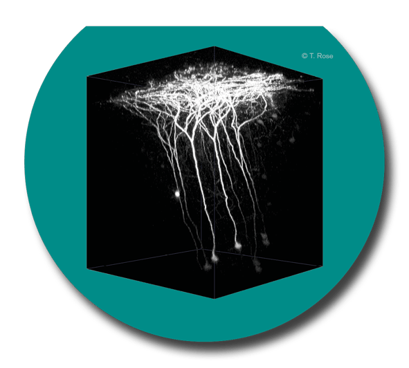
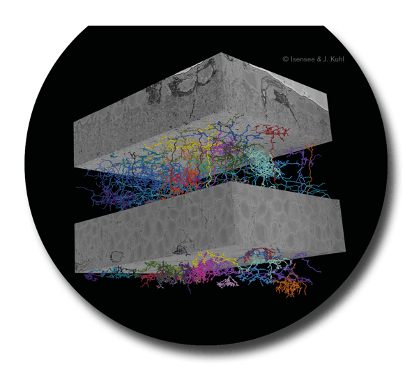
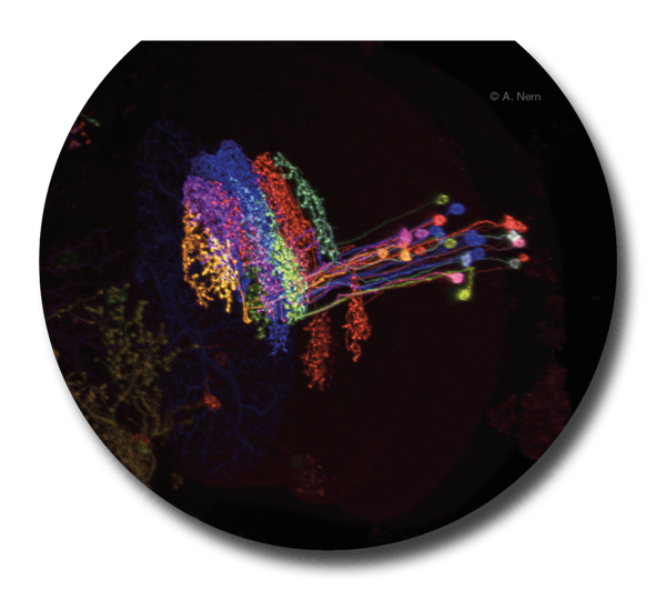
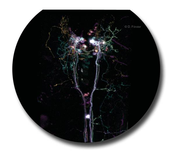
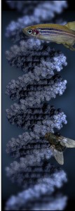
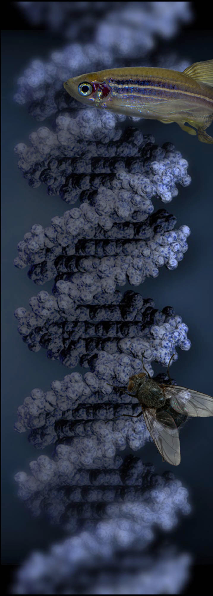
    Whenever possible, results are obtained using alternative methods to animal experiments. However, non-invasive procedures have mostly low resolution. Thus, the complex interactions of nerve cells with each other and their surroundings, as well as their influence on behavior can only be clarified with the aid of specially bred experimental animals. New methods and improved knowledge allow us today to gain important insights also from studies on flies, for example. No research at the Institutes is done on dogs, cats or monkeys.

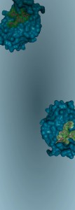
    In the 1960s, Albert Herz and his team explored the function and mechanisms of action of opium and opioids in mice and rats. The research team identified previously unknown opioids and found out where and how opioids work in the brain. They also showed how the investigated processes are influenced by drugs, stress and pain. The findings of these studies have become part of modern pain therapy (morphine), addiction therapy (methadone) and serve to assess the addiction potential of novel painkillers.

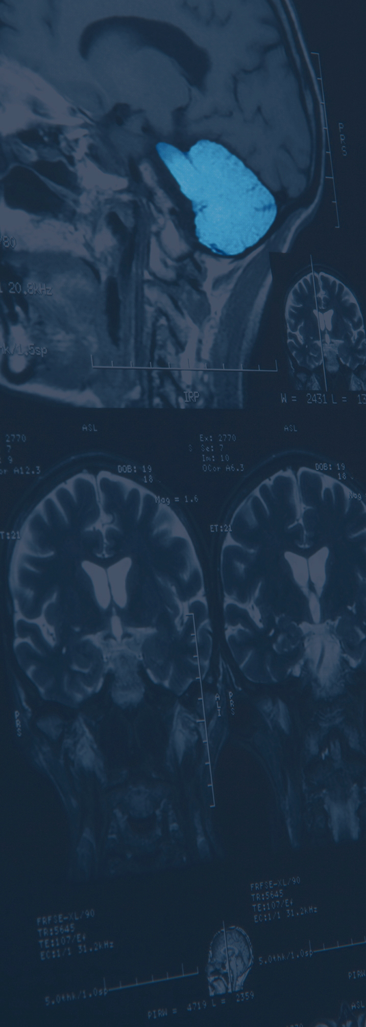
    Towards the end of the 1980s, Werner Risau, Axel Ullrich and colleagues at the Max Planck Institute for Biochemistry investigated growth factors that promote blood vessel formation. They found that small tumors produce a factor that stimulates the growth of blood vessels and thus promotes tumor growth. In the 1990s, the necessary animal experiments were carried out on mice in the USA. The end result was a highly effective cancer treatment (SUTENT®), launched in 2006.

    Hartmut Wekerle and his team investigate the fundamental causes of multiple sclerosis (MS), the most common autoimmune disease of the central nervous system. In the 1980s, they developed a rat model in which auto-aggressive T cells produce a MS-like disease. With the help of this animal model, Californian researchers were able to further elucidate mechanisms of the disease. This led to the approval of TYSABRI® in 2006, currently the most effective drug for relapsed MS.

    Are the results relevant to humans?

    Yes! Humans and animals are evolutionarily-related. As such, their genes, cells and organs often carry out similar or identical functions. Many diseases found in humans are also present in animals. Almost 90% of medications work equally well in humans as in pets. Approximately 70% of the undesirable effects of medications in humans can be predicted using appropriate animal models.

    Can animal experiments be avoided?

    In the planning and carrying out of animal experiments, the scientists act according to the 3Rs principle: Animal experiments are, if possible, to be avoided completely (Replacement), the number of animals used is the least possible (Reduction) and the animals’ suffering is kept to the absolute minimum (Refinement). In addition, researchers work on the development of alternative methods.

    Are people allowed to do that?

    Animal experiments can be associated with stress and suffering of animals. For this reason, before each animal experiment, researchers must carefully weigh up whether the expected gain in knowledge justifies the strain on the animals. This decision is never easy, but must be scientifically justified and must also withstand critical examination by an official ethical committee (§15 TierSchG) and a licensing authority.

    What does the law say?

    Experiments on vertebrates and cephalopods are subject to gaining prior approval (§8 TierSchG). For each animal experiment, an application for authorization must be submitted to the government. The application must set out the scientific background, the necessity, the suitability of the experimenter, the exact number of animals to be used and the degree of stress they will undergo. Approval will only be granted when the findings cannot be obtained by other means. Animal welfare officers are also involved in the approval process (§15 TierSchG). An approved experiment can be inspected at any time by the competent veterinary office without notice.

    What about the alternatives?

    Alternative methods are used whenever possible. Not only are they usually less complex and less expensive than animal experiments, they importantly avoid animal suffering. However, even in the age of computers, many of the very complex microscopic connections and processes can only be fully investigated in living organisms. As a result, many research questions cannot be answered without animal experiments.

    Where do the animals come from?

    Only healthy, unencumbered and suitable animals help research, because diseases and infections can alter results. The majority of animals used is bred in the Institutes’ animal facility. The quality and standard of the accommodation, breeding and care of the animals are under the constant scrutiny of veterinarians, animal keepers, animal welfare officers and the animal welfare committee of the Institutes.

  • Bringing new things to light

    Microscopes reveal minute details of the brain’s structure

    In order to truly understand the brain, it is imperative to visualize individual nerve cells, their connections and their activity. Thus, microscopy is one of the most important methods of neurobiological research.

    In the early 20th century, methods like the Nissl stain made it possible to investigate the distribution of nerve cells in sections of brain tissue under the light microscope.
    Late in the 20th and early in the 21st century, the development of fluorescent dyes labelling neural activity allowed the observation of individual cells’ activity and their components, as well as other cellular functions under the florescence microscope. However, to reach deeper brain areas, the development of the multiple-photon excitation microscope was necessary. This technique allows investigation not only of deep brain areas, but also for longer periods of time. Today, this microscopy technique, often in form of 2-photon fluorescence microscope, is an essential tool in neurobiological research.

    Apart from observing brain tissue, optogenetics now make it possible to use light to specifically activate or inhibit nerve cell activity. This enables, for example, live observations to determine which cells contribute to specific swimming or prey capture behaviors of zebrafish larvae. The latest development, the serial block face scanning electron microscope, allows reconstruction of an entire block of nervous tissue with all its nerve cells and their contact points in three dimensions – for the purpose of decoding the circuit diagram of the brain.

    Click for more details:

    Nissl bright-field microscopy


    Klappe 1

    2-photon fluorescent microscopy


    Klappe 3

    Serial Block Face Scanning Electron Microscopy


    Klappe 5

    Confocal fluorescent microscopy


    Klappe 2

    Optogenetic


    Klappe 4

Impressum